Here you can find an overview of our current research topics. There is also a list of awards of the visualization group available: go to awards page.
Research Meetings
- Narrative Visualization Meeting
- VR Meeting
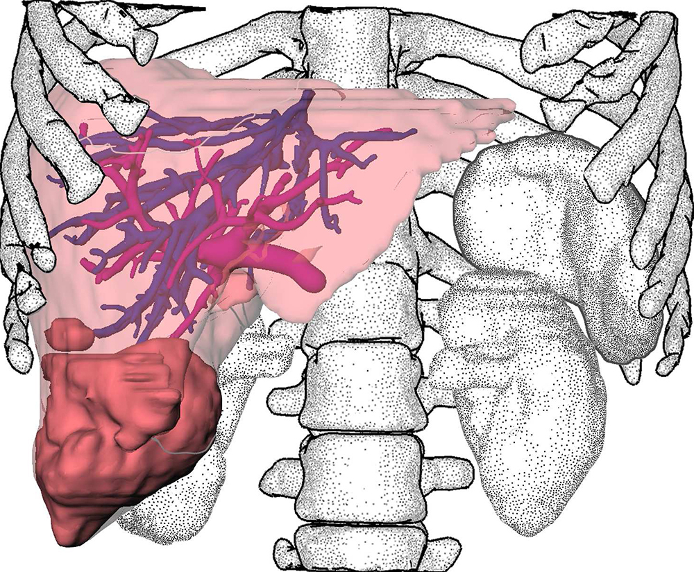
Preoperative Enhanced Analysis and Computational Optimization for Cholangiocarcinoma (PEACOCC)
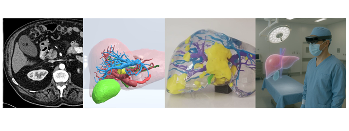
Effective preoperative planning is critical in oncologic surgery for perihilarcholangiocarcinoma (pCCA), given the high morbidity and mortality rates associatedwith these complex procedures and the considerable individual variability in liveranatomy. The PEACOCC project aims to enhance preoperative visualization by utilizingadvanced imaging techniques, specifically the latest photon-counting detectorcomputed tomography (PCD-CT), to improve the depiction of hilar and biliarystructures.
In addition, the project seeks to advance both surgical planning and intraoperativenavigation through the integration of virtual reality (VR), augmented reality (AR), and 3Dprinting technologies. While these tools have already demonstrated benefits in thetreatment of intrahepatic tumors, their application to pCCA remains underexplored andholds significant promise.
A key strength of the PEACOCC consortium lies in its well-established, interdisciplinarycollaboration between clinicians and engineers. This partnership combines extensiveexpertise in liver surgery, imaging, and computer-assisted analysis, positioning theproject to make impactful contributions to the field.
Narrative Medical Visualization
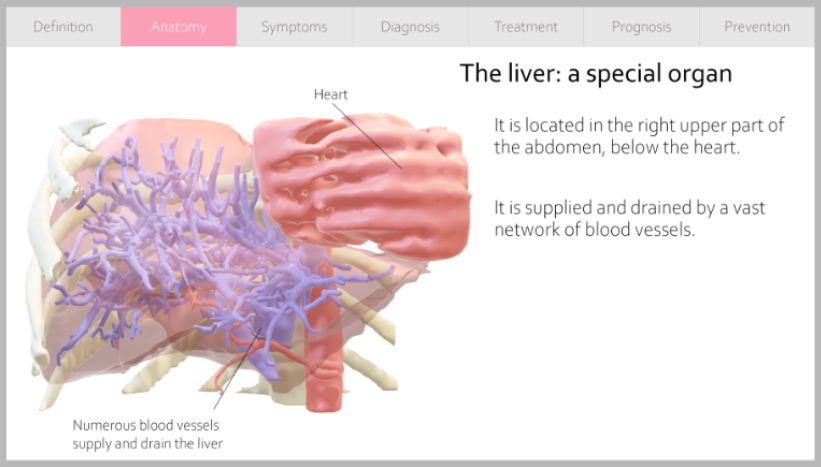
Medical visualization research has focused primarily on assisting physicians in diagnosis and treatment and, to a lesser extent, medical students, particularly in anatomy classes. However, medical data and research are also of interest to non-experts who lack detailed medical knowledge. They may be interested in links between lifestyle and disease, in understanding disease, and in (new) treatment options. Non-expert audiences require different design and narrative choices to create a clear, accessible, and understandable message. Interactive medical visualization aimed at this type of audience requires easier-to-understand displays than expert systems such as radiology workstations. Narrative visualization combines storytelling techniques with interactive graphics to communicate scientific results to a general audience.
Augmented Cooperation in Education and Training in Nuclear and Radiochemistry (A-CINCH)
EU – HORIZONT 2020; Project duration: 01.10.2020 to 30.09.2023
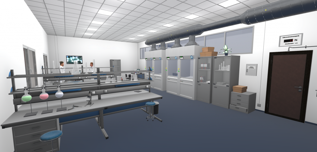
Expertise in nuclear and radiochemistry (NRC) is of strategic relevance in the nuclear energy sector and in many vital applications. The A-CINCH project primarily addresses the loss of the young generation’s interest for nuclear knowledge by focusing on secondary / high school students and teachers and involving them by the “Learn through Play” concept. This will be achieved by bringing advanced educational techniques such as state-of the art 3D virtual reality NRC laboratory, Massive Open Online Courses, RoboLab distance operated robotic experiments, Interactive Screen Experiments, NucWik database of teaching materials, or Flipped Classroom, into the NRC education. All the new and existing tools wrapped-up around the A-CINCH HUB – a user-friendly and easy-to-navigate single point of access – will contribute increasing the number of students and trainees in the field of nuclear and radiochemistry. Nuclear awareness will be further increased by the High School Teaching Package, Summer Schools for high school students, Teach the Teacher package and many others. Additionally, successful educational and training tools from previous projects will be continued and further developed. Networking is an important part of the project, facilitated by having ENEN as one of the partners and by having structural links with other Euratom projects, the EuChemS, the NRC-Network as well as by additional links with other end users and stakeholders including the high schools.
Development of Augmented and Virtual Multi-User Applications for Medical-Technical Exchange in Immersive Rooms (AVATAR)
Bund; 01.09.2018 to 31.08.2021
The exchange of surgical experience and competence nowadays mainly takes place at conferences, through the presentation of surgical videos and through the organisation of visits to each other. Complex manual skills and surgical techniques have to be newly developed, trained and passed on to younger surgeons or colleagues. With the methods currently used, this exchange is very costly and time-consuming.
In this project, VR interaction and visualization techniques will be developed to improve the exchange of experience and competence between medical professionals. In a virtual reality, several users are to train collaboratively – simultaneously and in real time. The positions of locally distributed persons will be determined using hybrid tracking systems based on ultra-wideband technologies and inertial sensors. On this basis, VR training scenarios are designed, implemented in a multi-user communication system and clinically evaluated over distance.
The innovation of this project is the combination of collaborative interaction and visualization techniques with hybrid tracking technologies in an advanced multi-user communication system. The project results should form a basis for the development of future VR-based communication and simulation systems in medicine.
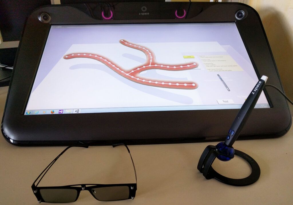
MEMoRIAL-M1.6 | Stent detection and enhancement
EU – ESF Sachsen-Anhalt; Project duration: 01.09.2017 to 30.04.2022
This projects aims at the
- automatic detection of stent and flow diverter markers,
- integration of stent deformation, as well as
- visualisation of the device s landing zone
to support the treatment of neurovascular diseases.
Stents and flow diverters are common devices for endovascular X-ray-guided treatment of neurovascular diseases such as aneurysms or artherosclerosis. Their visibility may, however, be hampered in clinical practice. To improve visibility especially during interventions, they are equipped with radiopaque markers. Given the limits of marker size, stents may, nevertheless, be almost invisible in fluoroscopy. Poor visibility of markers prompts physicians to spend more time on identifying the stent in fluoroscopy images, in turn leading to more time-consuming interventions and patients exposed to higher radiation doses.
This sub-project therefore addresses the detection of those markers in X-Ray images as well as the computer-based enhancement of their visibility. Furthermore, the 3D marker coordinates in space will be calculated using a second X-ray image shot from a different perspective and may provide additional information for the physician, e.g. revealing the stent deformation or landing zone of flow diverters.
Comparative analysis of the spatial and temporal progression of breast cancer lesions
EU – EFRE Sachsen-Anhalt; Project duration: 01.10.2019 to 30.09.2020
In the course of this project, a unique perfusion collective for the investigation of breast cancer lesions will be established. This collective will serve as a basis for a DACH application (joint DFG application with partners from Austria or Switzerland) on the topic of breast perfusion, between Magdeburg (OVGU) and Vienna (MUW). About 8-10 data sets are recorded per breast examination, which record the spread of contrast agent. Furthermore, patients come for follow-up examinations. This results in time-dependent data along two different scales/time axes, within an examination and between examinations.
The aim of this EFRE application is now to load these data and to display their temporal course within an examination. Furthermore, lesions will be segmented manually or semi-automatically for the determination of radiometric biomarkers. The EFRE funding period will be actively used to prepare the DACH application, to generate joint preliminary work and to prepare the data for radiomics and visual analytics of breast perfusion data. Looking ahead, prostate perfusion data (MUW) is still a possibility to be included in the DACH application, but the status of the data needs to be evaluated first.
Automatic segmentation of the aortic valve using deep learning
EU – EFRE Sachsen-Anhalt; Project duration: 01.04.2018 to 31.03.2020
The content of the project is to provide an automatic valve segmentation with features for manual post-processing in order to optimally support the physician in planning and performing surgery. The automatic valve segmentation will be performed using current methods of “deep learning”. According to the current state of research, these methods provide excellent results in the field of image segmentation. Quantifications of the valve geometry can be generated on a patient-specific basis after completion of the project. This allows for a more accurate and comprehensive characterization of the presenting clinical picture.
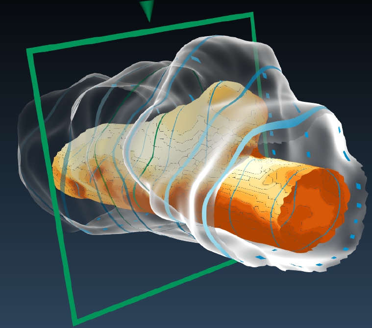
Qualitative and quantitative analysis of cardiac 4D PC-MRI blood flow data
Normwerterhebung etablierter Flussparameter bei einem gesunden Kollektiv und 1-Jahres Verlaufsevaluation ausgewählter Pathologien der Semilunarklappen mittels 4D PC-MRI
DFG; Project duration; 01.07.2016 to 30.06.2018
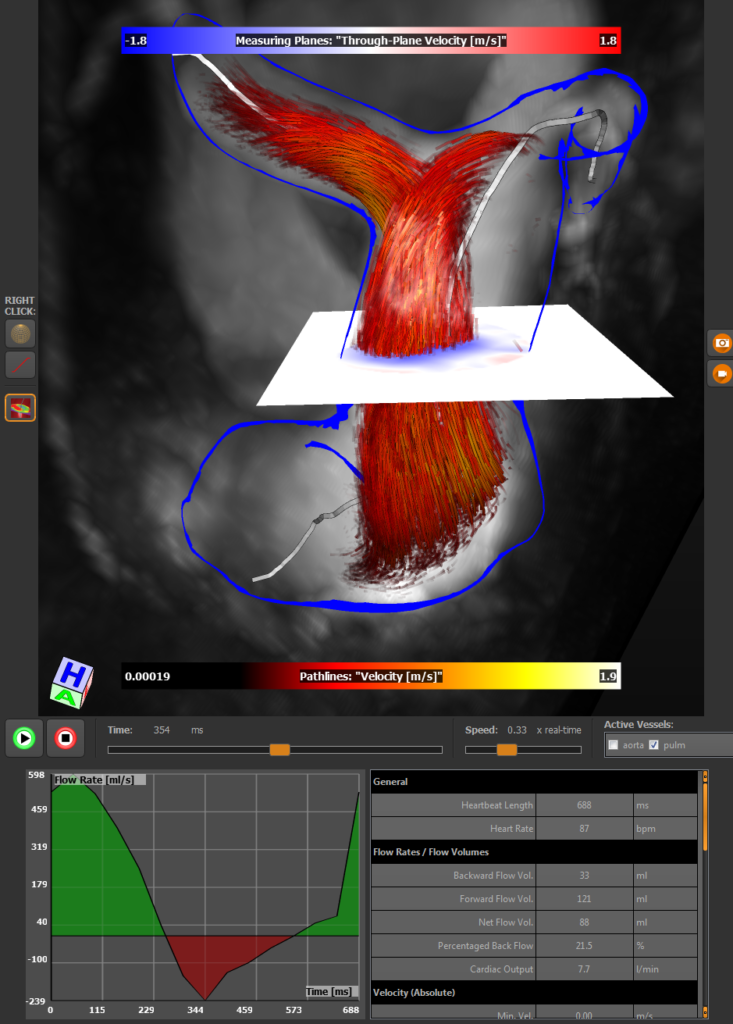
The genesis and progression of cardiovascular diseases (CVDs) depend on various factors. A better comprehension of patient-specific blood flow hemodynamics has great potential to increase their diagnosis, support treatment decision-making and provide a realistic forecast of such pathologies, facilitating a better implementation of preventative measures. Four-dimensional phase-contrast magnetic resonance imaging (4D PC-MRI) gained increasing importance and clinical attention in recent years. It is a non-invasive imaging modality that allows for time-resolved, three-dimensional measurement of blood flow information. The resulting 4D grid data, which contain vectors that represent the blood flow direction and velocity, are of limited spatio-temporal resolution and suffer from multiple artifacts, making complex image processing methods a prerequisite. Qualitative data analysis aims to depict the course of the blood flow with emphasis on specific flow patterns, such as vortex flow, which can be an indicator for different cardiovascular diseases. For this purpose, flow visualization techniques can be adapted to the cardiac context. Quantitative data analysis facilitates assessment of, e.g., the cardiac function by evaluating stroke volumes, heart valve performances by evaluating percentaged back flows, and fluid-vessel wall interactions by evaluating wall shear stress.
Virtual anatomy
BMWi/AIF; Project duration: 01.10.2015 to 31.03.2018
The “Virtual Anatomy” project develops and tests innovative concepts to modernize anatomy education. For example, the body donors that are used by medical students for preparation training are scanned in high resolution such that the preparation can also be simulated virtually. While the body donors can only be used for a limited time, the virtual models are basically always available. One of the practical research questions is how to directly integrate the viewing and analysis of 3D models into the curriculum of the anatomy education.
Home Training for the Treatment of Cognitive Disorders
EU – EFRE Sachsen-Anhalt; Project duration: 01.03.2017 to 28.02.2020
The cost pressure on rehabilitation hospitals results in stroke patients being released from hospital after 3-4 weeks and having further therapy with occupational therapists and neuropsychologists in private practice. However, under current conditions, the treatment intensity that is necessary for an efficient follow-up therapy is not further ensured after rehabilitation hospital discharge. To achieve therapeutic effects, the initiated therapy must be continued by intensive and preferably daily training.
This research project aims at the development of a system for the therapy of cognitive disorders for patients after stroke in home training. For this purpose, user interfaces with new interaction and visualization techniques shall be developed. Furthermore, studies shall validate whether reward and motivation techniques from computer games can be transferred to the new therapy software. For example, one element of the motivation and reward strategy is the suitable illustration of patient’s performance data.
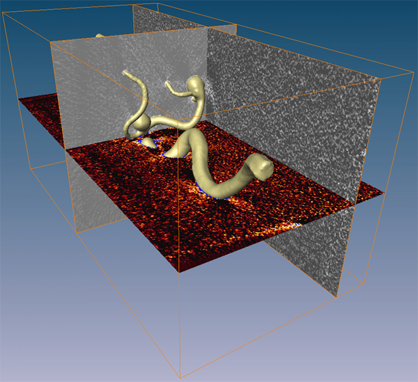
Perception-oriented blood flow visualization for patient-specific treatment of multiple aneurysms
EU – EFRE Sachsen-Anhalt; Project duration: 01.06.2016 to 31.05.2019
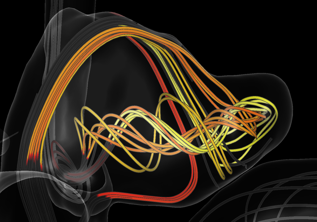
Cerebral aneurysms show a high prevalence in the western population, while their annual risk of rupture is below 1%. The bleeding caused by a rupture of such an aneurysms can have fatal consequences, but the treatment procedure itself is risky and can lead to severe complications.
Therefore, assessing the risk of rupture is vital to devise an optimal, patient-specific treatment plan – especially for patients with multiple aneurysms that may require multiple treatment sessions. The goal of this project is to develop and evaluate visualization techniques that support physicians with treatment decisions in such cases.
Visual Analytics in Public Health
DFG; Project duration 01.02.2012 to 31.12.2015
Unlike clinical applications, imaging in community medicine generates large amounts of imaging data from a large number of volunteers, with no specific question in mind for imaging. Analyses are typically performed on a large subject pool. Furthermore, such datasets can be analyzed over very long periods of time, so that analysis results should remain comparable with old studies. For this purpose, it must be guaranteed that the criteria according to which quantitative results are generated in the context of such an analysis are applied in the same way even after a longer period of time.
The aim of the overall project is to use visual analytics methods instead of many individual analysis methods for different questions in order to adapt a small pool of methods to the different questions using expert knowledge.
3D User Interfaces in medical therapy planning
Land (Sachsen-Anhalt); Project duration: 01.07.2014 to 30.09.2015
The dissertation deals with 3D user interfaces in medical therapy planning. Three aspects are examined in more detail:
- Interaction techniques
- input devices and
- output devices.
The goal of 3D user interfaces is to enable direct interaction with three-dimensional data through hardware. Here, the user can explore or manipulate 3D objects by e.g. pen- or gesture-based input. Stereoscopic and immersive output devices that convey motion parallax and binocular parallax, respectively, support depth perception in this process. Appropriate interaction techniques must be developed for the selected input and output devices. The application scenarios are chosen from medical therapy planning. It will be investigated how 3D user interfaces can improve or facilitate the planning of interventions.
Completed Topics
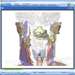
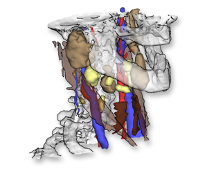
KOMET – Transfer platform in the field of medical technology: segmentation of soft tissue structures of the neck in MRI data
Land (Sachsen-Anhalt); Project duration: 01.10.2012 to 30.09.2013
In otorhinolaryngology, the diagnosis and therapy of malignant tumor diseases in the area of the mouth, nose and jaw represents an important area. The type of therapy depends on the overall assessment of the disease. Special care must be taken when resecting tumors or lymph nodes with metastases, as damage to nearby functional structures, such as large cervical vessels and cranial nerves, can lead to a significant reduction in the patient’s quality of life. Imaging techniques such as ultrasound, CT, MRI, or PET can be used to determine the malignancy of lymph nodes, for example, based on their size. However, size measurement is quite nonspecific and can lead to false positive or false negative findings. More specific is the detection of necrosis using MR imaging.
In this project, the techniques developed for CT data will be tested on MRI data and, if necessary, adapted so that surgical planning is also possible on this data. The expansion of surgical planning is motivated by the fact that MRI provides better soft tissue contrast and thus infiltration of at-risk structures can be better estimated. However, difficulties, e.g., due to inhomogeneities, geometric distortions, or different intensity values, are to be expected when analyzing MRI data. The adapted or newly developed image analysis and visualization techniques will finally be clinically evaluated.
KOMET – Transfer platform in the field of medical technology: development of a system for intuitive real-time exploration of three-dimensionally reconstructed endoscopy images
Land (Sachsen-Anhalt); Project duration: 01.10.2012 to 30.09.2013
Endoscopic examinations play an important role in the diagnosis of head and neck tumors because they provide information that complements tomographic imaging, especially with respect to tissue composition and surface structures. The project goal is to intuitively and efficiently visualize and explore the image data generated during an endoscopy, which can be reconstructed in a 3D model, using virtual endoscopy techniques. In this way, the examination results can be documented in a manner consistent with the nature of the examination. They are thus reproducible and can be reused in a variety of ways. The examiner can use the results as preparation for a surgical procedure, for patient education and for training. Telemedical examinations are also directly possible with them. In the event of a legal dispute, they help the physician to describe the planned procedure in a comprehensible manner.
The implementation of this goal requires the solution of some technically challenging tasks. In particular, the required real-time capability of three-dimensional, virtual exploration with the abundance of high-resolution data requires state-of-the-art visualization and interaction techniques. The reconstructed 3D model must be textured to a high quality so that the quality of the virtual exploration does not suffer. Since the surface does not have a regular shape, a largely distortion-free texture mapping is difficult.
Visualization of Cohort Study Data
Subproject of Visual Analytics in Public Health
DFG in SPP “Scalable Visual Analytics”
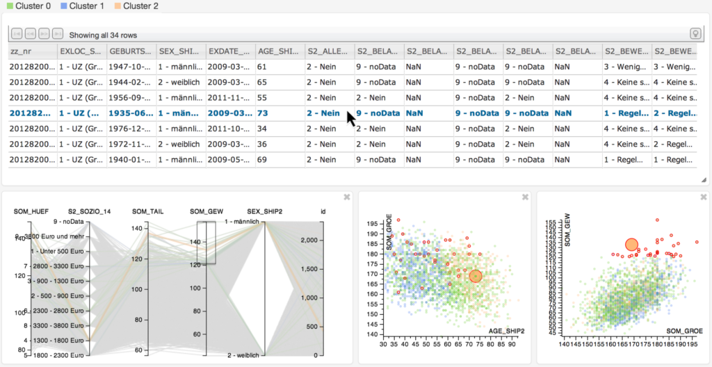
Large-scale longitudinal epidemiological studies investigate groups of people with common characteristics or experiences (a cohort) including socio-demographic and biological factors. Their goal is the characterization of health by identifying risk factors and their relations to diseases and the indication of a per subject risk of developing a disease. Carried out in waves over many years, they comprise thousands of individuals and ten thousands of variables. Cohort studies most recently also medical acquire image data. New Visual Analytics methods are developed as part of this project to support hypotheses validation and generation with respect to risk factors, inter-relations and relations A major goal is the visualization of inter- and intra-group statistical variance in organ shape.
Illustrative Visualization
In traditional illustrations, many well established techniques exist to communicate information. Their advantages consist in enhancing important information while unimportant information are suppressed. The aim of this project is to develop and evaluate such illustrative rendering techniques (like stippling, hatching, smart visibility). Our investigations are focused on applying these techniques in a medical context.
Perceptual Evaluations of Illustrative Techniques & Medical Visualizations

Many illustrative techniques for visualizing medical volume data and derived segmentation information have been developed and refined. However, it is difficult to decide which techniques should be used for particular applications, how they should be combined, which parameter adjustment achieve the best information transfer or how 2D and 3D displays influence the visualization perception and guide the users attention. This project investigates experimental studies to analyze illustrative visualization techniques used in 3D medical visualizations. We focus on the study design and try to adapt psychological theories and study concepts to complex 3D medical visualizations and techniques to analyze their benefit.
Interactive Visual Analysis of Toponome Data
In Toponomics, the function protein pattern in cells or tissue (the toponome) is imaged and analyzed for applications in toxicology, drug development and patient-drug-interaction. The most advanced imaging technique is robot-driven multi-parameter fluorescence microscopy. This technique is capable of co-mapping hundreds of proteins and their distribution and assembly in protein clusters across a cell or tissue sample. The imaging results in complex multi-parameter data composed of one slice or a 3D volume per protein affinity reagent. The goal of this project is to develop an interactive visual analysis framework which supports the biologist in evaluating the data.
Visualization & Exploration of Simulated and Measured Blood Flow Data
Subproject of Mobestan (Third party-project: state Saxony-Anhalt)
The aim of this project is to investigate the characteristics of blood flow in cerebral aneurysms. We focus on how new kinds endovasculare treatment can be used to influence the flow and decrease the risk for the patient. We deal with extraction, visualization and comparison of complex morphological information and flow features.
Visualization of Vessels
The research project for the visualization of vessels covers different research areas, e.g., the visualization of intravascular image data and model-based vessel visualization techniques. The intravascular imaging includes optical coherence tomography and intravascular ultrasound. The obtained medical image data provide valueable information about the vessel wall and its morphology which play an important role for diagnosis and evaluation of cardiovascular diseases like atherosclerotic plaque. Furthermore, special techniques to enable blood flow simulations and new projection-based techniques, that improve the conventional CPR views, were developed. Previous projects cover direct volume rendering techniques and transfer function adaptions as well as the vessel visualization with implicit surfaces.
Animation- & Exploration-Techniques on Geometric Models
In this project we develop facilities to create medical animations for intervention planning as well as for educational purposes. We also discuss the enhancement of interactive explorations with animations generated on the fly.
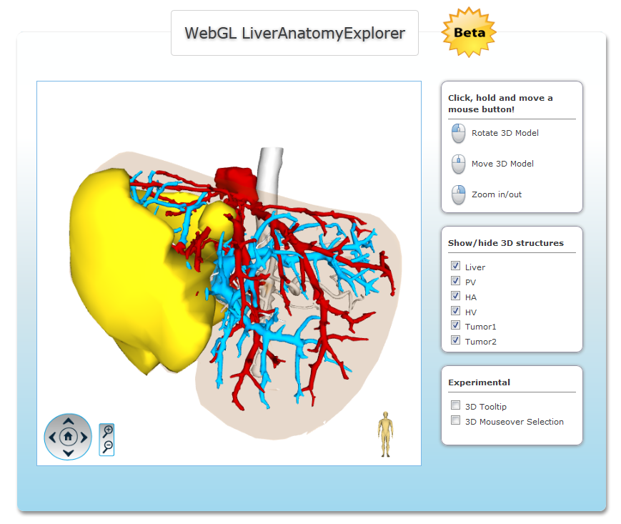
Computer-based Therapy Planning for Neck and Liver
Subproject of the DFG-Project VisHNO (DFG SPP 1124)
The aim of this project consists in developing methods for image analysis, visualization, and exploration of ENT and liver surgery planning. We focus on simulation of endoscopic intervention due to a user-driven navigation, usage of illustrative rendering techniques and model-based image analsysis.
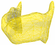
Generation of Surface Models from Segmented, Medical Image Data
Subproject of ViERforES (Third party-project: federal ministry of science and education)
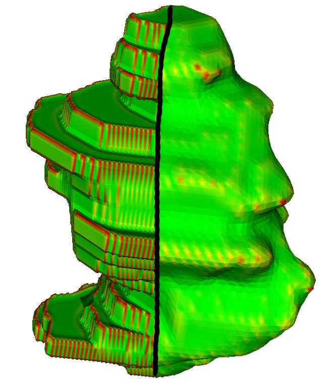
Surface models from medical image data typically suffer from artifacts such as staircases and terraces. Our goal is the (automated) generation of accurate and high quality surface models for surgical applications as well as simulations. The resulting surface models are evaluated regarding accuracy, smoothness, triangle quality (skewness), mesh resolution and homogeneity. The mesh generation process is focuses on image data preprocessing as well as postprocessing of the surface meshes (smoothing, remeshing, decimation).
Model-based Segmentation
Subproject of the DFG-Project VisHNO (DFG SPP 1124)
The efficient segmentation of anatomical and pathological structures in medical datasets is a prerequisite for various approaches to computer-supported therapy planning. This project is aimed at the model-based segmentation of anatomical structures from CT datasets using Stable 3D Mass-Spring Models (SMSM). Our long-term goal is to provide a general construction kit of elementary model-components which can be composed to build segmentation models for new structures easily.
Virtual Endoscopy
Supported via Priority Programme 1124, PR 660/3-1, PR 660/3-2 (Third party-project: DFG)
Minimally invasive endoscopic procedures are gaining importance, since they lead to a reduced trauma and hospitalization duration. Virtual endoscopy systems have been developed in order to simulate these interventions either for training purposes or for planning an actual intervention based on the CT or MRI data of the patient. We focus on interactive systems, that allow to interactively explore hollow space organs and support the patient education, operation planing or diagnosis of diseases. These tasks can be supported through the use real-time rendering techniques.
Visualization & Exploration of Perfusion Data
Subproject of Scalable Visual Analytics (DFG Research Priority Programme)
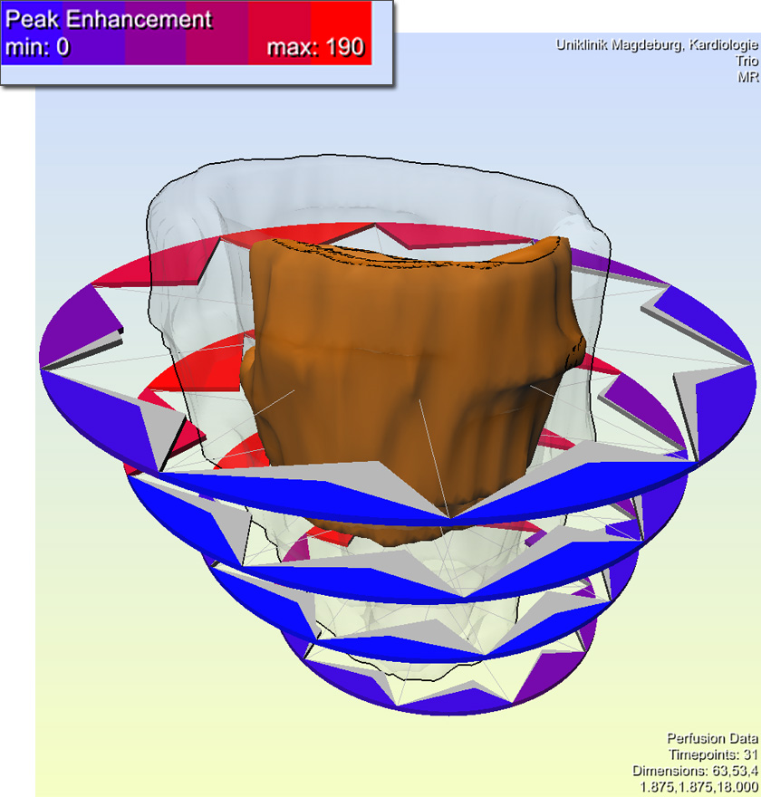
Perfusion data are dynamic medical image data which characterize the regional blood flow in tissue. We focus on the visualization and exploration of perfusion data for diagnostic purposes in three major application areas: ischemic stroke diagnosis, breast tumor diagnosis and the diagnosis of coronary heart disease.
Web Applications for Education and Training
Subproject of SurgeryNet (Third party-project: federal ministry of science and education)
The demand on education in surgery increases and classical educational methods can not meet it adequately. Therefore web-based training systems are developed, which have to be concepted and developed using several didactical and conceptual methods to allow learners more efficient learning. For procurement of explicit and implicit expertise and to represent the enormous anatomic diversity dynamic visualizations of the patients data are preferable. Modern web applications based on HTML 5 and WebGL are suitable for these requirements.