We present new surveys!
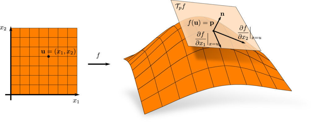
We submitted two surveys to the electronic archive arXiv. The first survey Feature Lines for Illustrating Medical Surface Models: Mathematical Background and Survey written by Kai Lawonn and Bernhard Preim is about feature lines. Here, several feature line methods are presented and analyzed. Furthermore, this survey provides also the discrete differential geometry background, which is necessary for understanding the feature line concepts.
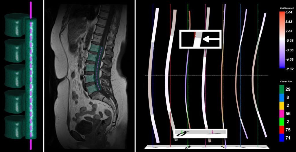
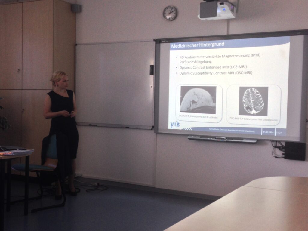
The second survey by Bernhard Preim et al. was a joint work with the visualization group of Bergen headed by Helwig Hauser, the image processing group headed by Klaus Tönnies, and Henry Völzke the head of the study of health in Pomerania. The survey Visual Analytics of Image-Centric Cohort Studies in Epidemiology discusses cohort studies and how they can be evaluated with visual analytics.
 We are proud that our article
  The Phd thesis of Kai Lawonn was selected the best Phd thesis of the Faculty in the academic year (1. 10. 2013 – 30. 9. 2014).
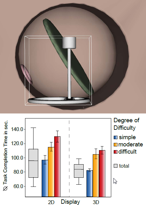
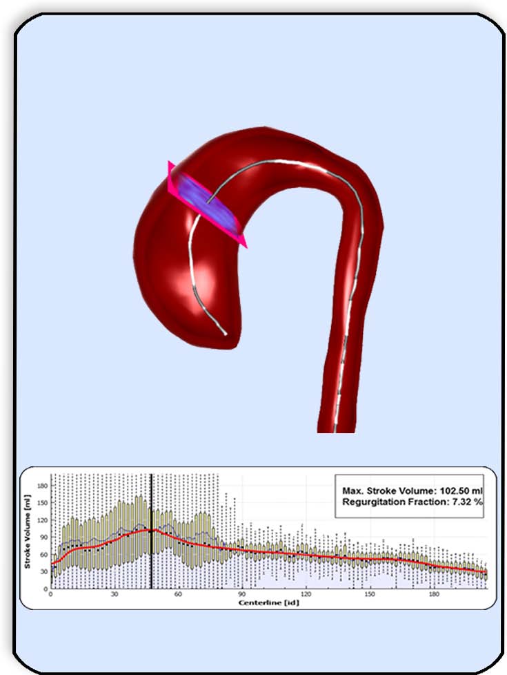
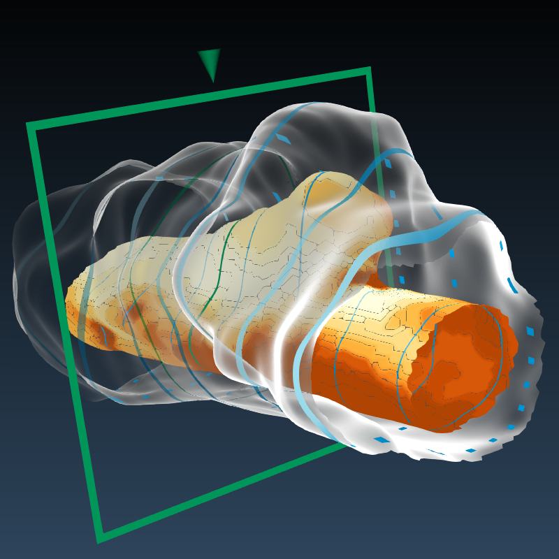
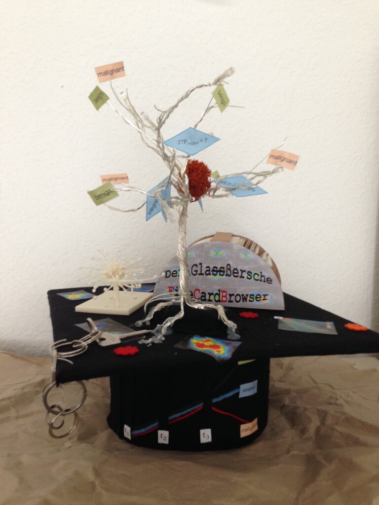
 Kai Lawonn has successfully defended his Phd thesis on “Illustrative Visualization of Medical Datasets” September, 15. Kai made a number of contributions related to feature lines applied to medical surface meshes. A systematic comparison of existing techniques, the development of new techniques, bridging between feature lines and hatchings, new applications in virtual endoscopy and molecular dynamics. Also for visualizing vascular surfaces along with internal blood flow Kai could strongly improve over existing methods. All this was accomplished in only 2.5 years. The Phd committee rated his performance as “Summa cum laude”.


 Tobias Mönch, Phd student of the visualization group, successfully defended his thesis “Context-Aware 3D Model Generation for Biomedical Applications”.