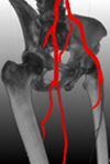
 Erkrankungen des Blutkreislaufs gehören zu den am weitesten verbreiteten Krankheiten in Industrienationen. So starben alleine in Deutschland im Jahr 2001 insgesamt 290.000 Personen an Erkrankungen des Herz-Kreislauf-Systems. Zuverlässige Diagnosesysteme und eine Verbesserung bestehender Verfahren sind daher von großer Bedeutung. Eine zentrale Aufgabe stellt hierbei eine geeignete Visualisierung des segmentierten Blutflußbaums beziehungsweise einzelner, erkrankter Abschnitte der Blutgefäße dar.
Diese Diplom-/Masterarbeit ist Teil eines Projekts zur vollautomatischen Segmentierung und Interpretation des arteriellen Gefäßbaums. Hierzu werden Verfahren entwickelt um unter Verwendung von anatomischen Hintergrundwissen, das in einem Referenzmodell abgelegt ist, den Gefäßbaum zu segmentieren. Anschließend soll die Diagnose durch geeignete Interpretation der Segmentierung optimal unterstützt werden. Die Grundlage des Systems sind dabei CT-Bilder, in denen Gefäße durch Kontrastmittel von umgebendem Gewebe hervorgehoben dargestellt werden.
Der Aufgabenbereich dieser Diplom-/Masterarbeit umfasst folgende Punkte:
- Effiziente Erzeugung eines Oberflächennetzes aus einem Graphen bestehend aus Gefäßmittelpunkten und zugehörigen Konturen
- 3D Visualisierung des Oberflächennetzes mit Hervorhebung der Abweichungen zwischen Segmentierung und Referenzmodell
- Visualisierung pathologischer Bereiche, wie Stenosen, Aneurysmen und Kalzifikationen
Voraussetzung für eine erfolgreiche Durchführung der Arbeit sind Kenntnisse der Computergraphik/Visualisierung sowie der Programmierung mit C++, wünschenswert ist außerdem Erfahrung in medizinischer Bildverarbeitung
Literatur: T. Beck, C. Biermann, D. Fritz et al.: “Robust model-based centerline extraction of vessels in CTA data”, Proc. SPIE, Vol. 7259, 2009
S. Grosskopf, C. Biermann, K. Deng et al.: “Accurate, fast, and robust vessel contour segmentation of CTA using and adaptive self-learning edge model”, Proc. SPIE, Vol. 7259, 2009

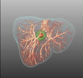 Chirurgische Eingriffe an der Leber, etwa zur Entfernung von Tumoren, gelten aufgrund der komplexen Struktur der Blutversorgung innerhalb der Leber als besonders schwierig. Für eine erfolgreiche Operation ist die genaue Kenntnis des Verlaufs der Blutgefäße von entscheidender Bedeutung, da sich an ihnen die Schnittführung der Resektion orientiert.
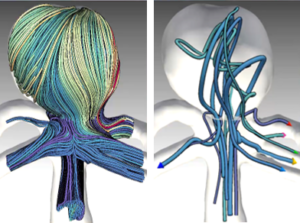 In cerebral aneurysm research, CFD simulations allow us to gain a better understanding of the dynamics of the blood flow. The simulated flow is often visualized using integral curves resulting in cluttered “spaghetti plots”. Advanced approaches group similar curves and show only selected representatives (image). These approaches however, fail in showing the clusters’ spatial extent. In this thesis, an interactive approach facilitating a continuous transition between the full set of integral curves and an uncluttered abstracted visualization shall be developed. Browsing back and forth through various levels of abstraction shall allow the user to grasp both, the general structure of the blood flow pattern as well as the spatial extent of individual substructures.
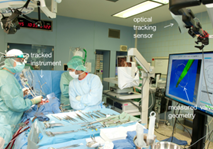 Im Rahmen der Arbeit sollen Stereoverfahren für die 3D-Rekonstruktion von Strukturen aus intraoperativen Endoskopiebildern entwickelt werden. Die Arbeit wird in enger Kooperation zwischen der Fakultät Informatik der OvGU Magdeburg (Dr. Sandy Engelhardt) und der Herzchirurgie des Universitätsklinikums Heidelberg (Prof. De Simone) durchgeführt. Weitere Themen für Abschlussarbeiten sind vorhanden. Melden Sie sich gern bei Interesse.
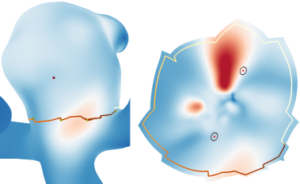 Zerebrale Aneurysmen sind pathologische Aussackungen der Gefäßwand, welche meistens an den Bifurkationen der großen Hirnarterien auftreten. Die Gefäßwand besitzt an diesen Stellen ein hohes Rupturisiko, was zu starken inneren Blutungen führt und in 60 % der Fälle den Tod des Patienten zur Folge hat. Daher ist eine patientenspezifische Einschätzung des Rupturrisikos nötig. Jedoch hängt die Ruptur von zahlreichen Kriterien ab, deren Zusammenhänge bisher nicht ausreichend verstanden sind. Blutflusssimulationen helfen dabei das patientenspezifische Rupturrisiko zu analysieren. Jedoch handelt es sich dabei um sehr komplexe Daten, was deren Auswertung enorm erschwert. Mit Hilfe von Standardtechniken wie Farbkodierungen und Animationen in 3D versuchen Experten rupturgefährdete Gefäßregionen ausfindig zu machen. Auftretende Verdeckungen machen es jedoch nahezu unmöglich über die Zeit Hochrisikoregionen zu finden. 2D Projektionen der 3D Gefäßgeometrie werden häufig eingesetzt, um verdeckungsfreie Überblicksvisualisierungen zu erzeugen. Jedoch führen derartige Projektionen zu Verzerrungen, die die Datenanalyse erschweren.