New Phd Student
 We are happy that Patrick Saalfeld got a grant for a Phd program from our local government to do research on 3D interaction related to treatment planning problems, such as implant placement and applicator positioning.
We are happy that Patrick Saalfeld got a grant for a Phd program from our local government to do research on 3D interaction related to treatment planning problems, such as implant placement and applicator positioning.
 Paolo Angelelli from the Visualization research group of the University of Bergen, Norway visited our group in October/November 2011. His visit initiated a long-term collaboration on investigating longitudinal medical and cohort studies between the Visualization group in Bergen (Paolo Angelelli, Cagatay Turkay, and Helwig Hauser), the Department of Biomedicine (Judit Haáasz, Erlend Hodneland and Arvid Lundervold) and the Department of Biological and Medical Psychology (Astri J. Lundervold) at the University of Bergen, and our Visualization group (Steffen Oeltze-Jafra, Bernhard Preim). In the paper resulting from this long-term effort, we demonstrate the interactive visual exploration and analysis of cohort study data, helping with the generation of new hypotheses and contributing to the process of validating them. We propose a data-cube based model which handles partially overlapping data subsets during the interactive visualization. We implemented this model in an application prototype, and used it to analyze data acquired in the context of a Norwegian cohort study on cognitive aging. A pre-print of the paper is available here: http://dx.doi.org/10.1109/MCG.2014.40.
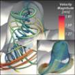 Flow data resulting from steady-state simulations of blood flow in cerebral aneurysms are generally visualized by a dense and cluttered set of streamlines. The paper describes a fully automatic approach for reducing visual clutter and exposing characteristic flow structures by clustering streamlines and computing cluster representatives. While individual clustering techniques have been applied before to streamlines in 3D flow fields, a general quantitative and a domain-specific qualitative evaluation of three state-of-the-art techniques are contributed. It is shown that clustering streamlines contributes to comparing and evaluating different virtual stenting strategies. The paper is the result of a collaboration between our Visualization group (Steffen Oeltze-Jafra, Bernhard Preim), the Institute of Fluid Dynamics and Thermodynamics of our University (Gábor Janiga), the Visual Computing group (Dirk J. Lehmann, Alexander Kuhn, Holger Theisel), and the Institute of Neuroradiology of our University Hospital (Oliver Beuing). It is available here: http://dx.doi.org/10.1109/TVCG.2013.2297914. The accompanying video can be found here: http://www.youtube.com/watch?v=-RVVgqDHzdc.
 Kai Lawonn has submitted his Phd thesis “Illustrative Visualization of Medical Data Sets” after a record time of 2 years and 4 months. As a mathematician, Kai soon become familiar with shader programming and advanced visualization techniques enabling him to make a number of strong contributions in illustrative and medical visualization. Among these contributions is a new method to generate smooth yet precise cutting lines based on an initial user-defined line, a substantial comparison of feature line techniques, the introduction of a new technique combining advantages of features lines and hatchings as well as improved smart visibility techniques to display vascular surfaces and embedded (simulated) blood flow. Also Kai was the first to display endoscopic views with feature lines and discussed pros and cons of their use.
Simon Adler, external Phd student of the visualization group, successfully defended his thesis on surgery simulation. Simon developed methods to efficiently cut in tetrahedral meshes, to deform organs and vascular structures.
 We are happy that two submissions of our group were accepted at EuroVis in Swansea (http://eurovis.swansea.ac.uk). The paper “Line Integral Convolution for Real-Time Illustration of Molecular Surface Shape and Salient Regions” was prepared by Kai Lawonn. It is a joint work with the Visualization group at University of Stuttgart where molecular dynamics simulations are carried out.
The paper “Comparative Blood Flow Visualization for Cerebral Aneurysm Treatment Assessment” is a result of our long-term effort to explore blood flow and to support neurovascular interventions. Rocco Gasteiger collaborated (again) with Roy van Pelt from Technical University of Eindhoven.
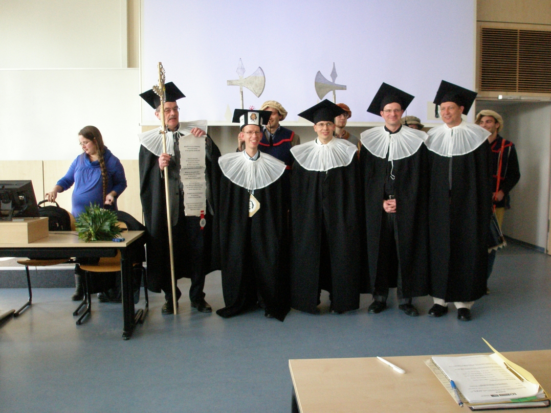
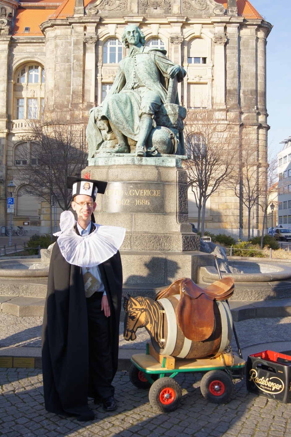
 Rocco Gasteiger successfully defended his PhD thesis on Visual Exploration of Cardiovascular Hemodynamics with summa cum laude at 07 February 2014. Rocco developed methods for expressive renderings of blood flow vessels with embedded flow information, to quantify the flow with respect to the inflow jet and the impingement zone in cerebral aneurysms as well as new exploration techniques, such as his FlowLens. During his PhD he collaborates very effectively with members from the group of Visual Computing, the Institute of Neuroradiology at the university hospital of Magdeburg, the Institute of Fluid Dynamics and Thermodynamics, the Department of Biomedical Magnetic Resonance as well as the Department of BioMedical Engineering at the Technical University of Eindhoven. Some visual impressions from the defense ceremony and party can be found here.
 Mathias Neugebauer submitted his PhD thesis „Computergestützte Exploration von zerebralen Aneurysmen – Geometrische Verarbeitung und interaktive Visualisierung“. In his work he describes various approaches for an anatomy-driven qualitative, visual exploration of flow in cerebral aneurysms. By reducing the spatial complexity and the usage of application-driven user interfaces and guided interaction, Mathias aims at bridging the gap between two expert domains: flow simulation and neuroradiology. Parts of this work were developed as part of MoBeStAn (“Modellierung und Beeinflussung von Strömung in zerebralen Aneurysmen”), a government-funded, interdisciplinary project.
 Tobias Mönch submitted his PhD thesis “Context-Aware 3D Model Generation for Biomedical Applications”. As a part of the BMBF-funded project “ViERforES”, Tobias analyzed the generation of anatomic surface models w.r.t. artifact reduction in the scope of characteristic medical applications, such as surgical planning and simulation, blood flow simulation, and rapid prototyping. By employing context knowledge on these specific applications and data, he developed new methods which improve the robustness of existing mesh smoothing filters and allow for a better preservation of the model quality.
Sylvia Glaßer submitted her PhD thesis “Visual Analysis, Clustering, and Classification of Contrast-Enhanced Tumor Perfusion MRI Data”. The thesis was realized as part of the DFG priority programme “Scaleable Visual Analytics”. Sylvia analyzed breast and brain tumor perfusion data combining automatic data analysis techniques and visual exploration for a more reliable discrimination of malignant and benign tumors.