Mathias Neugebauer submitted his PhD thesis „Computergestützte Exploration von zerebralen Aneurysmen – Geometrische Verarbeitung und interaktive Visualisierung“. In his work he describes various approaches for an anatomy-driven qualitative, visual exploration of flow in cerebral aneurysms. By reducing the spatial complexity and the usage of application-driven user interfaces and guided interaction, Mathias aims at bridging the gap between two expert domains: flow simulation and neuroradiology. Parts of this work were developed as part of MoBeStAn (“Modellierung und Beeinflussung von Strömung in zerebralen Aneurysmen”), a government-funded, interdisciplinary project.
Tobias Mönch submitted his PhD thesis “Context-Aware 3D Model Generation for Biomedical Applications”. As a part of the BMBF-funded project “ViERforES”, Tobias analyzed the generation of anatomic surface models w.r.t. artifact reduction in the scope of characteristic medical applications, such as surgical planning and simulation, blood flow simulation, and rapid prototyping. By employing context knowledge on these specific applications and data, he developed new methods which improve the robustness of existing mesh smoothing filters and allow for a better preservation of the model quality.
Sylvia Glaßer submitted her PhD thesis “Visual Analysis, Clustering, and Classification of Contrast-Enhanced Tumor Perfusion MRI Data”. The thesis was realized as part of the DFG priority programme “Scaleable Visual Analytics”. Sylvia analyzed breast and brain tumor perfusion data combining automatic data analysis techniques and visual exploration for a more reliable discrimination of malignant and benign tumors.
The second edition of the book “Visualization in Medicine” has appeared at Elsevier. The slightly changed title “Visual Computing in Medicine” reflects that image analysis, human-computer interaction and simulation play a greater role. The whole content is completely updated. In addition to the printed book, five chapters are available freely online.
The paper “Adapted Spectral Clustering for Evaluation and Classification of DCE-MRI Breast Tumors” by Sylvia Glaßer and colleagues introduces a spectral clustering approach for breast tumor data. The clustering parameters are automatically derived such that the clustering results can be employed for an automatic classification into benign and malignant breast lesions.
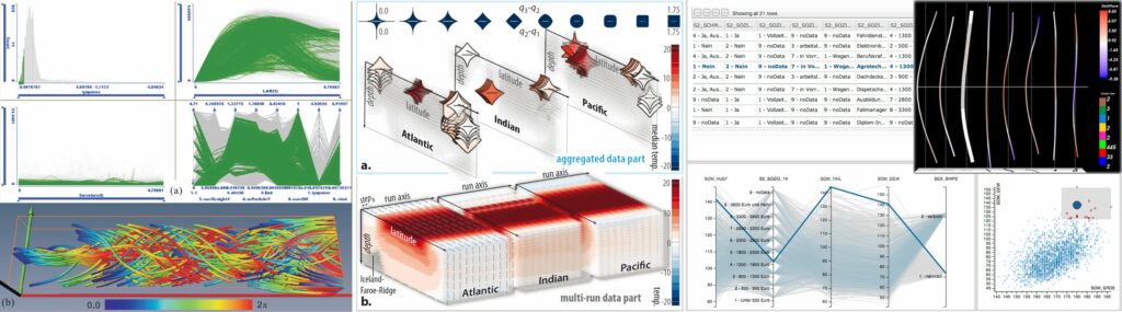
The paper “Socio-demographic and Medical Attribute Data in Cohort Studies” by Paul Klemm and colleagues proposes an alternative approach for analyzing significant interactions for identifying risk factors in epidemiological cohort study data by incorporating clustering algorithms with a Visual Analytics system to form subject groups. This is the basis for an exploratory analysis of the underlying parameter interactions. With the presented system, groups can be automatically determined to provide insights into this complex data.
The paper “Illustrative Visualization of Endoscopic Views” was prepared by Kai Lawonn and colleagues and deals with the application of illustrative line renderings on endoscopic views. Different line drawing concepts were examined and the ability to represent interior branches as well as specific anatomic features was assessed.
Rocco Gasteiger submitted his Phd thesis today. Starting in 2007 and sometimes side-tracked by other projects he had to take care of, Rocco dealt with the exploration of measured and simulated blood flow data, comparing results, developing new exploration techniques, such as his FlowLens. In a close cooperation with Roy van Pelt and Anna Vilanova , Rocco developed techniques to quantify the flow with respect to the inflow jet and the impingement zone. Besides his two most essential papers that appeared at IEEE TVCG, another contribution of Rocco is extraordinary: a new approach to depict the vascular morphology and internal flow, published at the VCBM Eurographics Workshop in 2010 and being effectively the most heavily cited paper.
We are happy that our tutorial submission entitled “Interactive Visual Analysis of Scientific Data” has been accepted as a half-day tutorial for VisWeek 2013. The tutorial is a collaborative effort of Helwig Hauser from the University of Bergen, Norway, Johannes Kehrer from the Vienna University of Technology, Austria, and Steffen Oeltze (organizer). The tutorial has been held once before at VisWeek 2012. This year, the tutorial has been significantly changed, e.g., to present new application fields of Interactive Visual Analysis (IVA) to the audience: IVA of epidemiological and flow data.
We are happy that the two submissions for the SciVis conference within IEEE VisWeek (http://ieeevis.org/) were accepted for presentation.
The paper “Semi-Automatic Vortex Extraction in 4D PC-MRI Cardiac Blood Flow Data using Line Predicates” was prepared by Ben Köhler and colleagues and represents the major results of Ben’s Master thesis. Features of measured cardiac blood flow are related to severe pathologies. This work was only possible with substantial support from the Herzzentrum Leipzig (Prof. Gutberlet, Dr. Uta Preim) who provided these very special kind of data and greatly helped to interpret them and guide the analysis towards clinically relevant features.
Jan Kretschmar (SIEMENS Forchheim and Phd student in Erlangen) prepared a paper “Interactive Patient-Specific Vascular Modeling with Sweep Surfaces” on the creation of vascular surfaces models that are not only smooth and accurate but also appropriate for blood flow simulation. We could slightly contribute to Jan’s work and extend the cooperation that already lead to a EuroVis paper last year.
After the presentation at VCBM in Norrköping 2012, Tobias Mönch was invited to prepare an enhanced article for the Computer Graphics Forum on the framework for interactive mesh smoothing, primarily for anatomical surface models. After careful reviewing, the paper was now accepted and appeared online http://onlinelibrary.wiley.com/doi/10.1111/cgf.12165/abstract.
It represents the highlight of Tobias’ upcoming Phd thesis. The paper was joint work with Kai Lawonn, Christoph Kubisch (nVidia) and Rüdiger Westermann (TU Munich) who contributed to algorithmic descriptions and efficient hardware implementation.
 Mathias Neugebauer submitted his PhD thesis „Computergestützte Exploration von zerebralen Aneurysmen – Geometrische Verarbeitung und interaktive Visualisierung“. In his work he describes various approaches for an anatomy-driven qualitative, visual exploration of flow in cerebral aneurysms. By reducing the spatial complexity and the usage of application-driven user interfaces and guided interaction, Mathias aims at bridging the gap between two expert domains: flow simulation and neuroradiology. Parts of this work were developed as part of MoBeStAn (“Modellierung und Beeinflussung von Strömung in zerebralen Aneurysmen”), a government-funded, interdisciplinary project.
 Tobias Mönch submitted his PhD thesis “Context-Aware 3D Model Generation for Biomedical Applications”. As a part of the BMBF-funded project “ViERforES”, Tobias analyzed the generation of anatomic surface models w.r.t. artifact reduction in the scope of characteristic medical applications, such as surgical planning and simulation, blood flow simulation, and rapid prototyping. By employing context knowledge on these specific applications and data, he developed new methods which improve the robustness of existing mesh smoothing filters and allow for a better preservation of the model quality.
 The second edition of the book “Visualization in Medicine” has appeared at Elsevier. The slightly changed title “Visual Computing in Medicine” reflects that image analysis, human-computer interaction and simulation play a greater role. The whole content is completely updated. In addition to the printed book, five chapters are
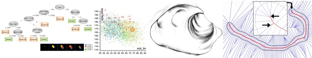
 Rocco Gasteiger submitted his Phd thesis today. Starting in 2007 and sometimes side-tracked by other projects he had to take care of, Rocco dealt with the exploration of measured and simulated blood flow data, comparing results, developing new exploration techniques, such as his FlowLens. In a close cooperation with
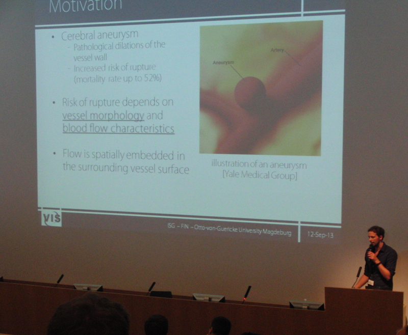
 We are happy that the two submissions for the SciVis conference within IEEE VisWeek (