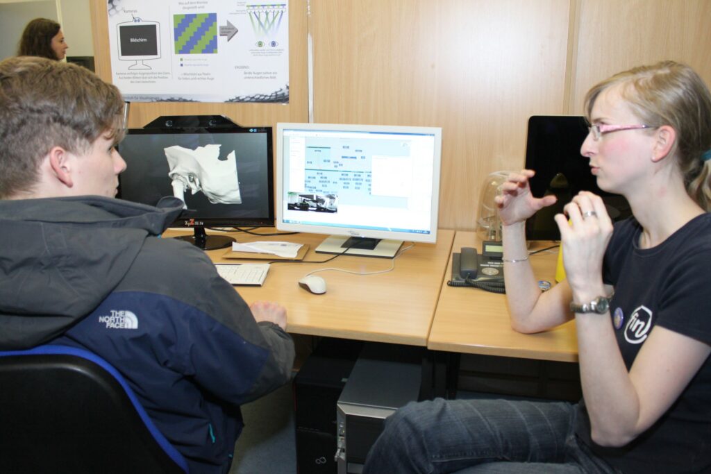
Around 1300 visitors attended the “Long Night of Science” on June 1st, 2013 at the Faculty of Computer Science. Not only on June 1st, but also during the Campus Days (held on May, 31 and June, 1st), the Visualization group presented current research work on “Medical 3D Visualization and Interaction for Therapy Planning”.
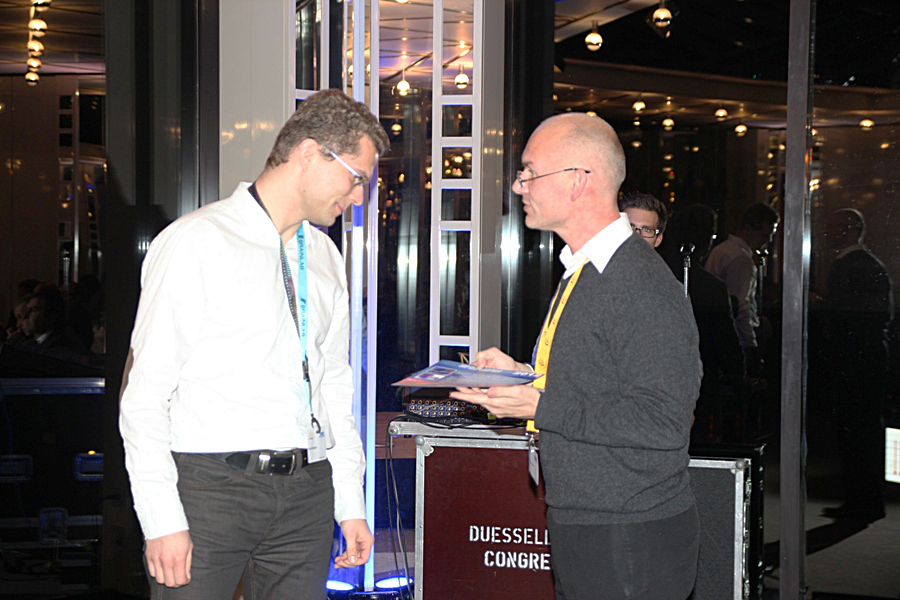
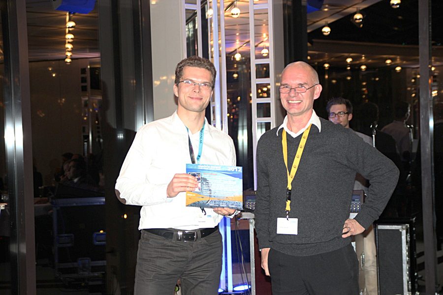
We are happy to announce that members of the visualization group were awarded with the 2nd prize of the Dirk Bartz Prize for Visual Computing in Medicine 2013. The entry was titled “Interactive Visual Exploration of Hemodynamics in Cerebral Aneurysms” and it summarizes the work of Mathias Neugebauer, Rocco Gasteiger, Gabor Janiga (ISUT), Oliver Beuing (Radiology Dept. University Hospital), and Bernhard Preim. The award ceremony was part of this year’s EuroGraphics Award ceremony, in the beautiful Teatre Municipal de Girona (Girona, Spain).
Our submission “The LiverAnatomyExplorer: A WebGL-based Surgical Teaching Tool” was accepted for a special issue of IEEE Computer Graphics and Applications on Web-Based 3D visualizations.
The paper describes the design, implementation and evaluation of a liver anatomy system that conveys different variants of the liver anatomy based on 13 datasets. 2D slice-based and 3D visualizations with a number of interaction techniques to explore the data are provided. State-of-the-art Web3D technologies and frameworks, like WebGL and X3DOM, are employed to provide a solution without any plugins. In contrast to other web-based training systems, the LiverAnatomyExplorer is based on a representative set of cases and provides more interactive features. The paper was authored by Steven Birr, Jeanette Mönch, Uta and Bernhard Preim.
We are happy that two of our submissions for the EuroVis conference in Leipzig were finally accepted and will appear in the Computer Graphics Forum.
Kai Lawonn is the principal author of the paper “Streamlines for Illustrative Real-time Rendering” where he suggests a new method to place lines on surfaces of complex (anatomical) surface models. These lines are seeded along curvature-related vector fields and provide more shape information than conventional feature lines. The line generation process, including curvature estimation, as well as the efficient rendering is described in the paper.
Mathias Neugebauer introduced “AmniVis – A System for Qualitative Exploration of Near-Wall Hemodynamics in Cerebral Aneurysms”. The paper is based on many in-depth discussions with neuroradiologists and aims to present results of unsteady (time-dependent) CFD simulations intuitively and effectively. Essential questions, guiding this research, are: How can the user efficiently select relevant time steps? What features are particularly interesting? How they can be emphasized and assessed?
Die Arbeitsgruppe “Medizinische Visualisierung” an der Universität Magdeburg feiert ihr 10-jähriges Bestehen. Aus diesem Anlass wird es am 26. April 2013 ab 15 Uhr im Hörsaal der Fakultät für Informatik (G29-307) ein Symposium geben, bei dem ein kleiner Einblick in die vergangenen 10 Jahre und in die Arbeiten der Arbeitsgruppe, ehemaliger Mitarbeiter und eng verbundener Partner gegeben wird.
We are looking forward to the visit of Anders Ynnerman, clearly the key visualization researcher in Sweden and in particular the leading medical visualization expert there. He is known among others for his work on interactive exploration of large medical data using modern surface and touch technology, see his popular TED talk.
Anders leads the famous visualization center in Norrköping, closely collaborates with the imaging center in Linköping and recently co-organized the Eurographics Workshop on Visual Computing in Biology in Medicine. Anders visits both, the visual computing and the visualization group and gives a talk in the visual computing colloquium (Link: http://wwwisg.cs.uni-magdeburg.de/visual/index.php?article_id=163) series on 1:15 pm on Friday early afternoon.
Our journal submission “The LiverSurgeryTrainer – Training of Computer-Based Planning in Liver Surgery” was accepted at the International Journal of Computer Assisted Radiology and Surgery. The paper with Jeanette Mönch as Principal author documents as a summary our long-term efforts to create a software system for case-based training, including expert opinions, intraoperative videos and resection proposals. Surgeons may assess the operability and resectability and compare their decisions with expert decisions and their explanations. Many thanks for the cooperation to Fraunhofer MEVIS, in particular Christian Hansen and Prof. Karl Oldhafer, our major medical partner. We are also thankful to many other surgeons for providing the necessary feedback.
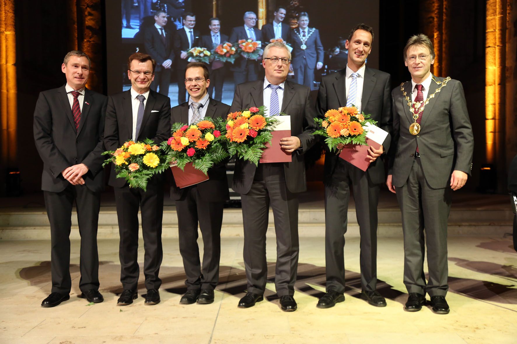
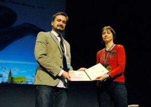
Rocco Gasteiger’s work on the visual exploration of cerebral blood flow was selected for the first price from 17 submissions to this year’s Karl-Heinz Höhne Award. At the photos, Dr. Stefan Zachow from Zuse-Institute Berlin who was the head of the selection committee (together with Prof. Dorit Merhof), awards the price. Rocco was the first author of two related IEEE TVCG papers exploring features of cerebral blood flow in the presence of cerebral aneuryms.
Besides his numerous own ideas, concepts, and implementations, he carefully collaborated with colleagues and other groups. He integrated important works from our partners in flow simulation (Gabor Janiga, Dominique Thevenin), from our medical partners (Oliver Beuing, Martin Skalej), our friends from Eindhoven (Roy van Pelt, Anna Vilanova) as well our colleagues from the visual computing group (Dirk Lehmann, Holger Theisel) and finally, Rocco very effectively teamed up with Mathias in our group for many years now.
 We are happy to announce that members of the visualization group were awarded with the 2nd prize of the Dirk Bartz Prize for Visual Computing in Medicine 2013. The entry was titled “Interactive Visual Exploration of Hemodynamics in Cerebral Aneurysms” and it summarizes the work of Mathias Neugebauer, Rocco Gasteiger, Gabor Janiga (ISUT), Oliver Beuing (Radiology Dept. University Hospital), and Bernhard Preim. The award ceremony was part of this year’s EuroGraphics Award ceremony, in the beautiful Teatre Municipal de Girona (Girona, Spain).
 Our submission “The LiverAnatomyExplorer: A WebGL-based Surgical Teaching Tool” was accepted for a special issue of IEEE Computer Graphics and Applications on Web-Based 3D visualizations.
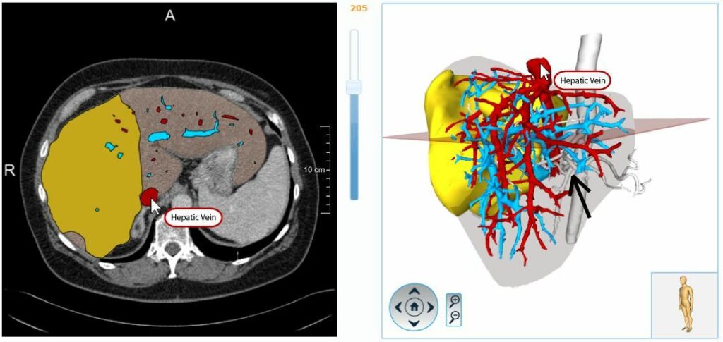
 Kai Lawonn is the principal author of the paper “Streamlines for Illustrative Real-time Rendering” where he suggests a new method to place lines on surfaces of complex (anatomical) surface models. These lines are seeded along curvature-related vector fields and provide more shape information than conventional feature lines. The line generation process, including curvature estimation, as well as the efficient rendering is described in the paper.
 Mathias Neugebauer introduced “AmniVis – A System for Qualitative Exploration of Near-Wall Hemodynamics in Cerebral Aneurysms”. The paper is based on many in-depth discussions with neuroradiologists and aims to present results of unsteady (time-dependent) CFD simulations intuitively and effectively. Essential questions, guiding this research, are: How can the user efficiently select relevant time steps? What features are particularly interesting? How they can be emphasized and assessed?

 On January 30, the kick-off event for the research campus STIMULATE (Solution Centre for Image guided local Therapies) takes place, see the
  We are looking forward to the visit of Anders Ynnerman, clearly the key visualization researcher in Sweden and in particular the leading medical visualization expert there. He is known among others for his work on interactive exploration of large medical data using modern surface and touch technology, see his popular
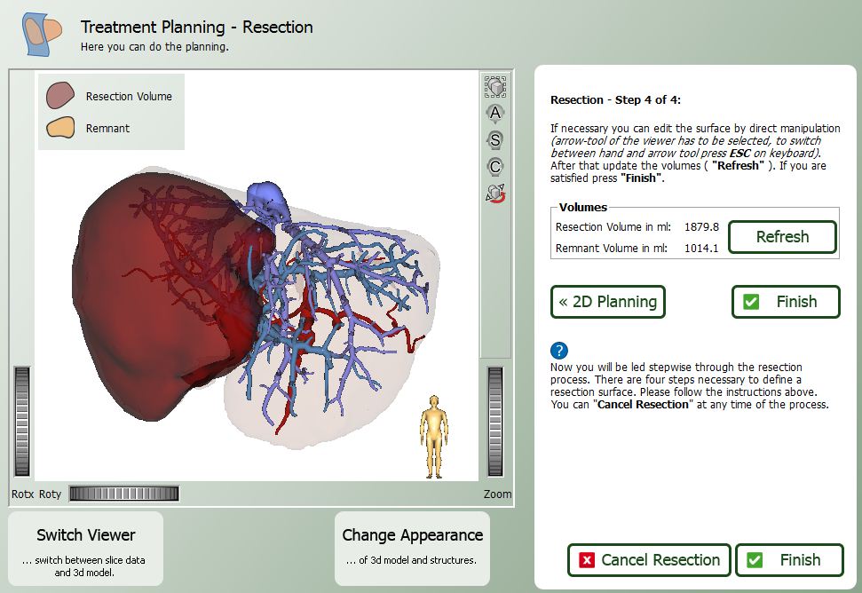
 Our journal submission “The LiverSurgeryTrainer – Training of Computer-Based Planning in Liver Surgery” was accepted at the International Journal of Computer Assisted Radiology and Surgery. The paper with Jeanette Mönch as Principal author documents as a summary our long-term efforts to create a software system for case-based training, including expert opinions, intraoperative videos and resection proposals. Surgeons may assess the operability and resectability and compare their decisions with expert decisions and their explanations. Many thanks for the cooperation to Fraunhofer MEVIS, in particular Christian Hansen and Prof. Karl Oldhafer, our major medical partner. We are also thankful to many other surgeons for providing the necessary feedback.