Vis-Group at EuroVis 2013
We are happy that two of our submissions for the EuroVis conference in Leipzig were finally accepted and will appear in the Computer Graphics Forum.
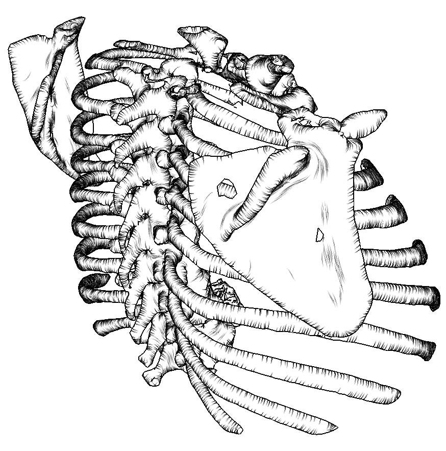
 Kai Lawonn is the principal author of the paper “Streamlines for Illustrative Real-time Rendering” where he suggests a new method to place lines on surfaces of complex (anatomical) surface models. These lines are seeded along curvature-related vector fields and provide more shape information than conventional feature lines. The line generation process, including curvature estimation, as well as the efficient rendering is described in the paper.
 Mathias Neugebauer introduced “AmniVis – A System for Qualitative Exploration of Near-Wall Hemodynamics in Cerebral Aneurysms”. The paper is based on many in-depth discussions with neuroradiologists and aims to present results of unsteady (time-dependent) CFD simulations intuitively and effectively. Essential questions, guiding this research, are: How can the user efficiently select relevant time steps? What features are particularly interesting? How they can be emphasized and assessed?

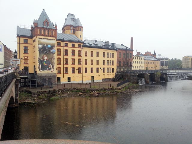
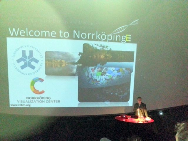
 On January 30, the kick-off event for the research campus STIMULATE (Solution Centre for Image guided local Therapies) takes place, see the
  We are looking forward to the visit of Anders Ynnerman, clearly the key visualization researcher in Sweden and in particular the leading medical visualization expert there. He is known among others for his work on interactive exploration of large medical data using modern surface and touch technology, see his popular
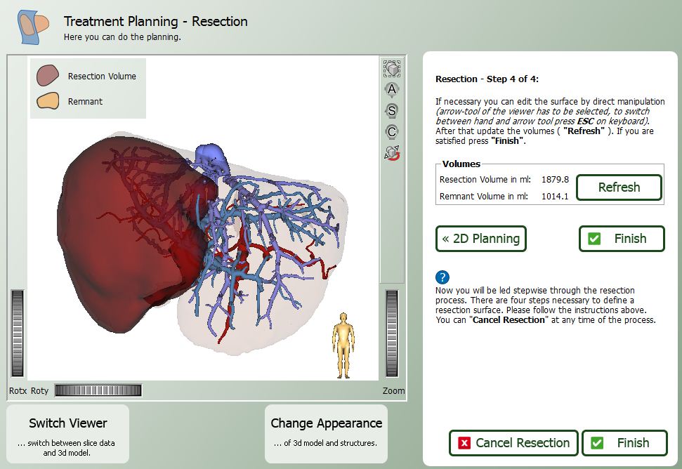
 Our journal submission “The LiverSurgeryTrainer – Training of Computer-Based Planning in Liver Surgery” was accepted at the International Journal of Computer Assisted Radiology and Surgery. The paper with Jeanette Mönch as Principal author documents as a summary our long-term efforts to create a software system for case-based training, including expert opinions, intraoperative videos and resection proposals. Surgeons may assess the operability and resectability and compare their decisions with expert decisions and their explanations. Many thanks for the cooperation to Fraunhofer MEVIS, in particular Christian Hansen and Prof. Karl Oldhafer, our major medical partner. We are also thankful to many other surgeons for providing the necessary feedback.
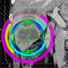
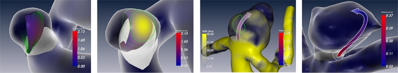
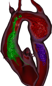 Ben developed tools to process these time-dependent blood flow data, searching for topologically interesting regions, like those with high helicality and vorticity. The visual exploration of blood flow in the human Aorta is then focussed on such regions.